Can useful phase and absorbance information be recovered when an X-ray hologram contains very few photons? In our 2026 study, we developed MorpHoloNet-X, a physics-driven neural network for single-shot reconstruction under weak illumination. The approach combines sample-specific prior knowledge with an explicit model of X-ray wave propagation.
Why weak illumination makes reconstruction difficult
X-ray holographic microscopy records interference between the incident beam and waves affected by a specimen. The detector measures intensity, while phase and absorbance must be inferred. With limited photon counts, shot noise obscures interference fringes and can distort the recovered physical quantities.
Phase describes how the specimen changes the wave’s phase; absorbance describes attenuation. Recovering both provides information beyond the apparent brightness of a reconstructed image.
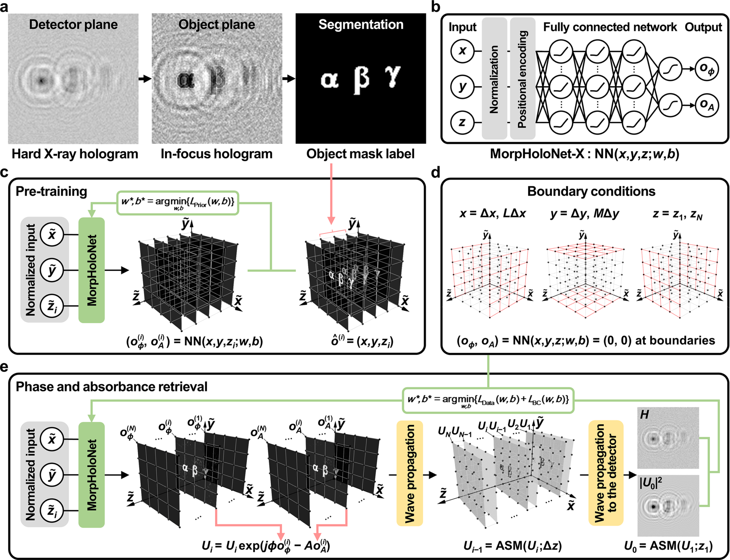
Method: a neural field constrained by imaging physics
- Identify a candidate object region. An in-focus reconstruction is segmented to obtain an object mask. Approximate specimen depth and available characterization guide the reconstruction.
- Represent the object in three dimensions. The network receives spatial coordinates and predicts separate voxel-wise object values for phase and absorbance.
- Pre-train using prior knowledge. Object masks near the expected depth initialize the candidate region. Approximate optical properties provide initial phase and absorbance coefficients.
- Simulate the measured hologram. The angular spectrum method propagates a complex wavefield through the volume, incorporating phase shifts and attenuation. Boundary constraints suppress objects at the volume edges.
- Optimize against the measurement. The model minimizes the mean squared difference between simulated detector intensity and the recorded hologram. Early stopping is needed to avoid fitting shot noise.
This is a reconstruction optimized for the input hologram using physics and specimen priors. “Single-shot” refers to the hologram used for reconstruction; it does not mean that sample characterization, calibration or computational optimization are unnecessary.
Experimental setup
| Parameter | Reported setup |
|---|---|
| Facility | 7C X-ray Nano Imaging beamline, PLS-II |
| X-ray energy | 9.344 keV |
| Waveguide channel | 140 × 120 nm |
| Waveguide-to-object distance | 8 mm |
| Waveguide-to-detector distance | 15 cm |
| Object-plane pixel size | 17.15 nm |
| Exposure time | 10 s |
| Lateral resolution by Fourier ring correlation | 213 nm |
Pixel size and measured resolution are different: the 17.15 nm sampling interval does not establish 17.15 nm resolving power. The reported setup achieved 213 nm lateral resolution by Fourier ring correlation.
What the validation showed
The study compared MorpHoloNet-X with the Gerchberg–Saxton algorithm and a residual U-Net using synthetic shot-noise-limited holograms and an experimental gold resolution target. In the experimental target, the reference phase shift was 0.278 rad.
| Method | Recovered phase shift (rad) | Phase error | Absorbance error |
|---|---|---|---|
| Gerchberg–Saxton | 0.066 ± 0.033 | 76.2% | 50.7% |
| Residual U-Net | 0.129 ± 0.018 | 53.7% | 7.2% |
| MorpHoloNet-X | 0.240 ± 0.027 | 13.5% | 9.7% |
MorpHoloNet-X substantially improved experimental phase retrieval. Residual U-Net had the smaller mean absorbance error in this experiment, whereas MorpHoloNet-X better preserved high-frequency edge features. The results therefore should not be described as uniform superiority across every measurement.
Why it matters, and what remains unresolved
The contribution is a way to incorporate wave physics and sample knowledge when hardware limits photon availability. It offers a potential route toward imaging with weak illumination or shorter acquisitions, but the reported experimental exposure was 10 seconds.
The method depends on object masks and approximate optical properties. Severe shot noise still degraded reconstruction; excessive optimization could overfit noise. The forward model also used plane-wave propagation despite cone-beam illumination, a simplification that may contribute to error. Full experimental validation of single-shot 3D morphology reconstruction under the present shot-noise-limited conditions remains a future step.
For a complementary example of how synchrotron measurement conditions affect structural interpretation, read our lithium adsorption and synchrotron XRD research note. Diffraction and holographic microscopy answer different questions, but both require attention to signal quality and acquisition conditions.
Original motivation and future direction
An early motivation for this work was to explore whether energy-dependent X-ray absorption could be combined with rapid depth reconstruction to visualize processes inside lithium-ion batteries. Measurements near the absorption edges of elements such as nickel and manganese could potentially provide information about their spatial distribution and chemical states. Tracking these changes during battery operation might help reveal how reactions progress unevenly within electrode materials.
This was a longer-term direction rather than an application demonstrated in the present study. Here, we focused on recovering phase and absorbance from X-ray holograms under weak illumination at a fixed energy. We hoped that improving the reliability of these reconstructions could provide a foundation for future energy-resolved, three-dimensional imaging of battery reactions.
Figure credit: Kim et al. (2026), Figure 1, Journal of Synchrotron Radiation. Original paper. Reproduced without modification under CC BY 4.0.
Original paper
Kim J, Lim J, Jo S, Park S, Lee SJ. Phase and absorbance retrieval in X-ray holographic microscopy under weak illumination using physics-driven neural networks. Journal of Synchrotron Radiation 33, 794–805 (2026). Read the original paper. Methods: Sections 2.1 and 5; experimental comparison: Figure 7; limitations: Section 3. The original article is published under CC BY 4.0.
Keywords: X-ray holographic microscopy; phase retrieval; absorbance retrieval; MorpHoloNet-X; physics-driven neural networks; synchrotron imaging.