Before a baby takes its first breath, blood already has a route around the lungs. The placenta supplies oxygen, and the foramen ovale, a normal opening between the atria, allows part of the blood entering the right atrium to pass into the left atrium. From there, it can continue through the left ventricle and into the aorta.
That route is familiar from fetal circulation diagrams. Seeing it inside a living fetal heart is a different challenge. In this case report, our team used Doppler ultrasound-gated 4D flow MRI to visualize the pathway in a healthy fetus at 32 weeks of gestation. The scan took 3 minutes and 37 seconds, without contrast agents or anesthesia.
A small opening with a big role
The fetal circulation works differently from the circulation after birth. Because oxygen comes from the placenta, the lungs do not yet perform gas exchange. The foramen ovale helps direct placental blood toward the left side of the heart and the systemic circulation.
Fetal echocardiography remains the primary imaging method and can assess flow across this opening. The additional question for 4D flow MRI is spatial: can we follow the route through the heart in three dimensions, rather than observe it only within a selected ultrasound view?
How do you synchronize MRI with a fetal heartbeat?
The fetal heart is small, fast, and moving. In this case, the heart rate was approximately 140 beats per minute. Synchronizing MRI with that cycle is difficult because conventional surface ECG gating is not readily available for the fetus.
A Doppler ultrasound probe placed on the maternal abdomen detected fetal cardiac motion and supplied a timing signal to the MRI scanner. This allowed a standard vendor-provided Cartesian 4D flow sequence to collect cardiac-synchronized data during maternal free breathing. The images were reconstructed on the scanner, making image-quality assessment possible immediately after acquisition.
The distinction matters: this workflow avoided specialized offline image reconstruction, but it still required post-processing. Velocity data were preprocessed in MATLAB, anatomy was segmented in 3D Slicer, and particle traces were visualized in EnSight.
Following the route, one stage at a time
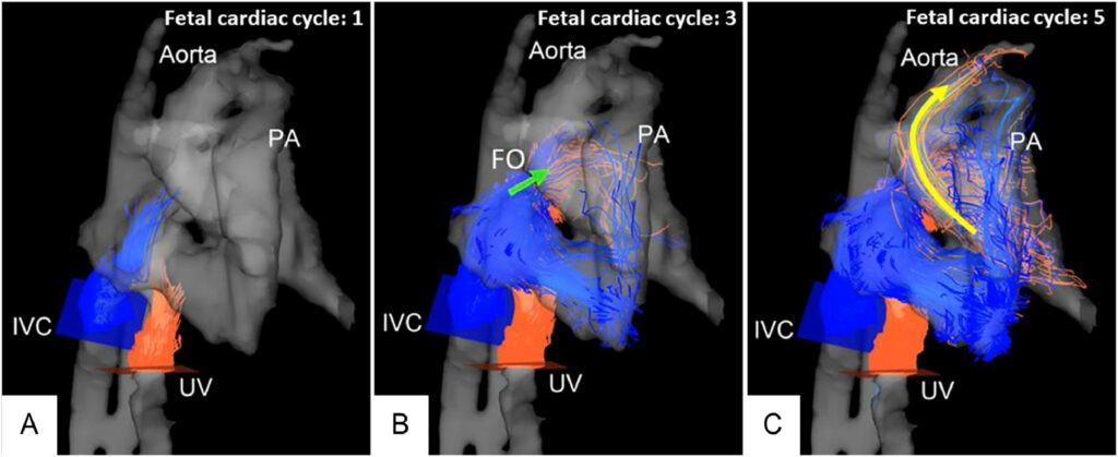
Figure 1 turns an anatomical diagram into a flow visualization. Particle traces seeded at the umbilical vein and inferior vena cava were integrated across multiple cardiac cycles:
- Entering the heart: the traces enter the right atrium and approach the atrial septum.
- Crossing the shortcut: right-to-left flow across the foramen ovale becomes visible.
- Continuing outward: traces that crossed into the left atrium continue toward the left ventricular outflow tract and aorta.
These are computational trajectories derived from MRI velocity measurements, not individually labeled blood cells. Their colors identify the locations where traces were seeded; they are not direct measurements of oxygen saturation. The visualization nevertheless provides an intuitive view of how the fetal circulation connects.
What this case demonstrates
The main result is feasibility of visualizing normal foramen ovale flow in vivo using a short acquisition, Doppler gating, and on-scanner reconstruction. Flow in the ductus venosus and ductus arteriosus was also identified. The pregnancy continued without complication, and the infant was delivered at term with normal postnatal adaptation.
This was a visualization report in one healthy fetus. It did not validate quantitative foramen ovale shunt volumes, establish diagnostic thresholds, or show improved clinical outcomes. A coherent pathline image also does not independently prove the accuracy of every velocity measurement.
Why the next step is interesting
If the pathway can be visualized consistently, a future question is how much blood crosses the opening and whether its direction or distribution changes in congenital heart disease. Retrospective analysis planes within a 4D flow volume could potentially support measurements of forward, reverse, and net flow.
Those possibilities still need validation. Small intracardiac structures, limited spatial and temporal resolution, signal quality, fetal motion, and an unstable Doppler window can all affect the result. Earlier gestation, unusual fetal position, and arrhythmias add further challenges. Interpolation makes the displayed image grid finer but does not replace the acquired resolution.
The appeal of this report is therefore practical as well as physiological: it shows a recognizable fetal blood-flow pathway with a workflow that combines a conventional MRI sequence and external Doppler timing. Larger studies will need to establish how reliably it works across patients and whether quantitative measurements add useful information to fetal echocardiography.
Acquisition at a glance
| Gestational age | 32 weeks |
|---|---|
| Scanner | 3 T Philips Ingenia, software R5.6; 32-channel torso coil |
| Gating | Doppler ultrasound, smart-sync (Northh Medical) |
| Acquired voxel size | 2.5 × 2.5 × 2.5 mm³ |
| Reconstructed voxel size | 1.04 × 1.04 × 2.5 mm³ |
| Temporal resolution | 50 ms |
| VENC | 150 cm/s |
| TE / TR | 1.98 / 3.25 ms |
| Flip angle / acceleration | 6° / SENSE 3 |
| Acquisition time | 3 minutes 37 seconds |
Original paper
Gerstner Saucedo J, Fujiwara T, Englund EK, Park S, Friesen R, Browne LP, Barker AJ. Following the flow: in vivo imaging of fetal foramen ovale physiology using four-dimensional flow magnetic resonance imaging with doppler ultrasound gating. Radiology Case Reports. 2026;21:564–569. Published online November 14, 2025. Read the original paper.
Keywords: fetal 4D flow MRI, foramen ovale, fetal circulation, Doppler ultrasound gating, fetal cardiac MRI, intracardiac shunting, pathline visualization.